Mitoribosome Assembly
The Problem.
Prof. Alexey Amunts (Stockholm University) needed an illustration to depict and promote his lab's new research uncovering a mechanism of assembly of the human mitoribosome small subunit. The objective was to create visuals that communicated the core discovery of the paper in a way that would be accessible and engaging to a broad audience. Continue below for the case-study of the design process, written by BK SciViz owner, Brad Krajina.
An Initial Solution.
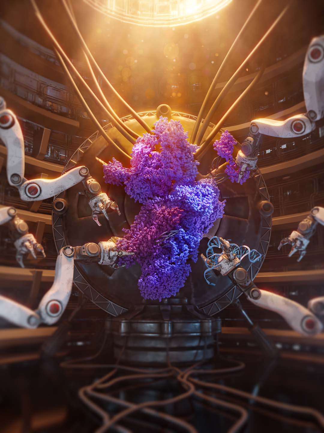
I pitched an initial idea to Prof. Amunts: The Mitoribosome being "assembled" in a sci-fi silo, with robot arms bringing the individual sub-components together. All of the molecular structures were generated using the electron microscopy data generated by Alexey's lab.
Refining the Design.
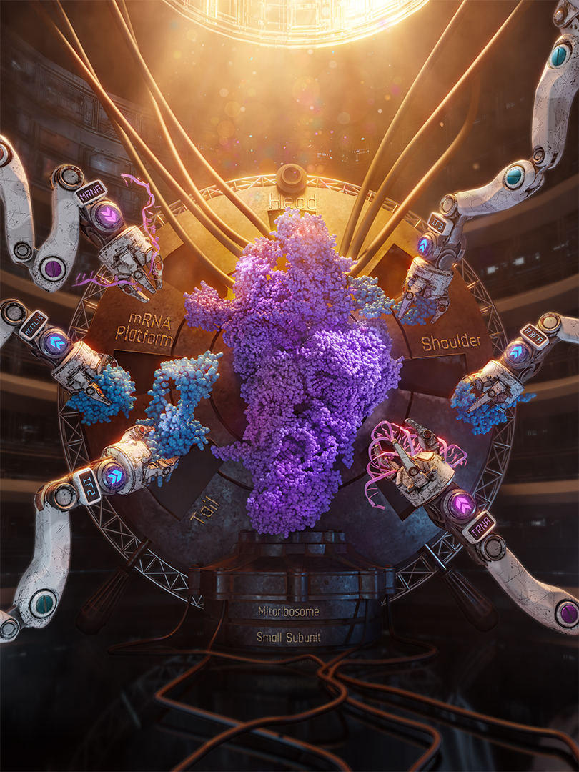
Prof. Amunts liked the idea and the design, but he wanted to bring more of the details of the study into the design. His idea was to change the design of the robot arms so that each one clearly identified the components that were involved, and also to clearly show which components were being added to the structure, and which were being removed.

Bringing the Assembly to Life.
After we finished the illustration, Prof. Amunts reached out to see if we could transform the illustration into a short movie to capture the dynamics described in their study. We put together a plan for the shots, I developed a rough cut animation, and we finalized the movie that would be used to help promote their new research.
The Outcome.
By translating complex molecular ideas into engaging visuals, this work helped Prof. Amunts bring visibility to his innovative research. The animation and illustration collectively garnered over 50,000 organic views on social media, driving over 250 users to follow the link to the online publication.
Contact
info@bksciviz.com
Email us directly for a free consultation.